Categories Products for Mass Spectrometry In-vitro SILAC Proteomics Media and Kits
In-vitro SILAC Proteomics Media and Kits
In vitro SILAC (Stable Isotope Labelling of Amino Acids in Cell Culture) has been proven a powerful technique for quantitative proteomics in cell culture. The method is robust and provides accurate results.[1]
Silantes provides
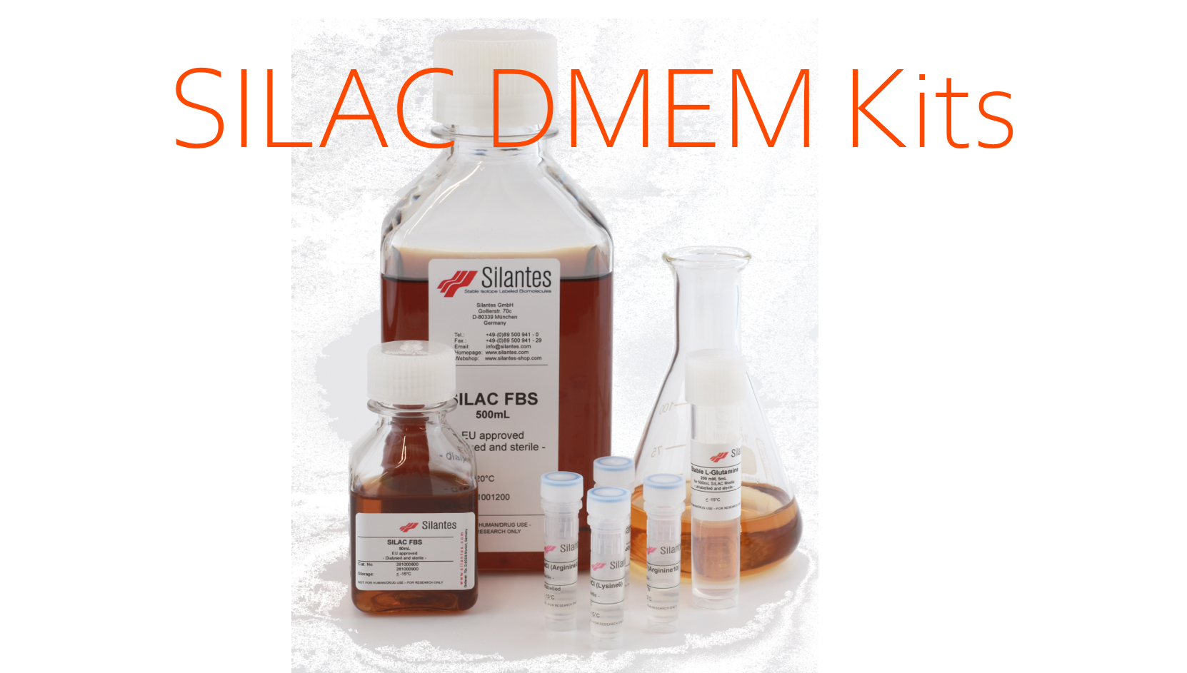
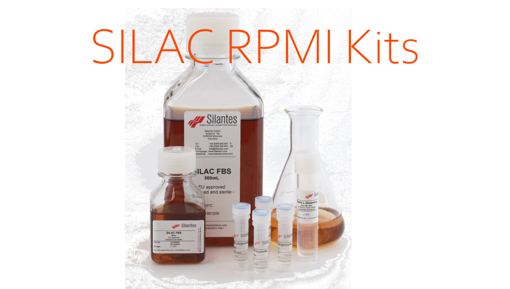
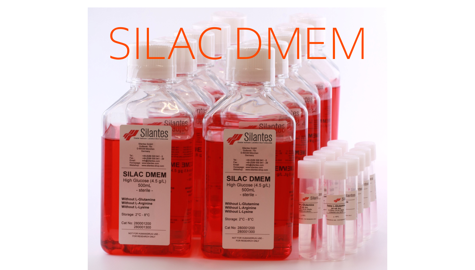
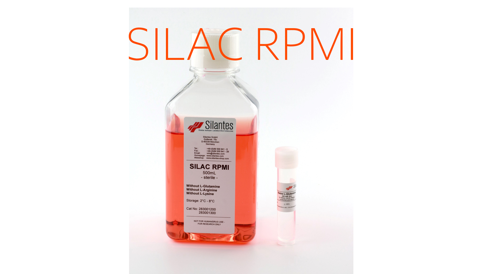
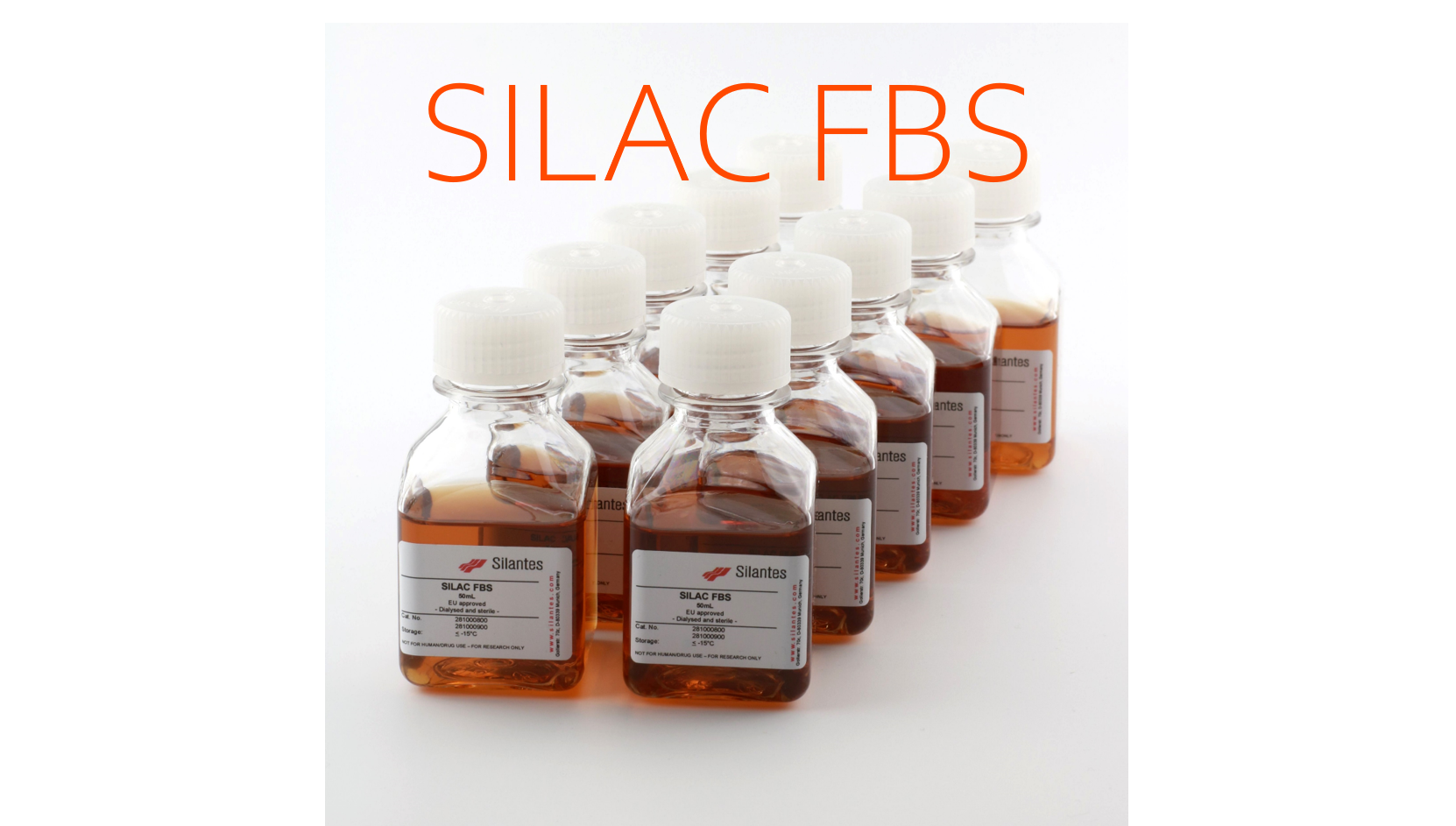
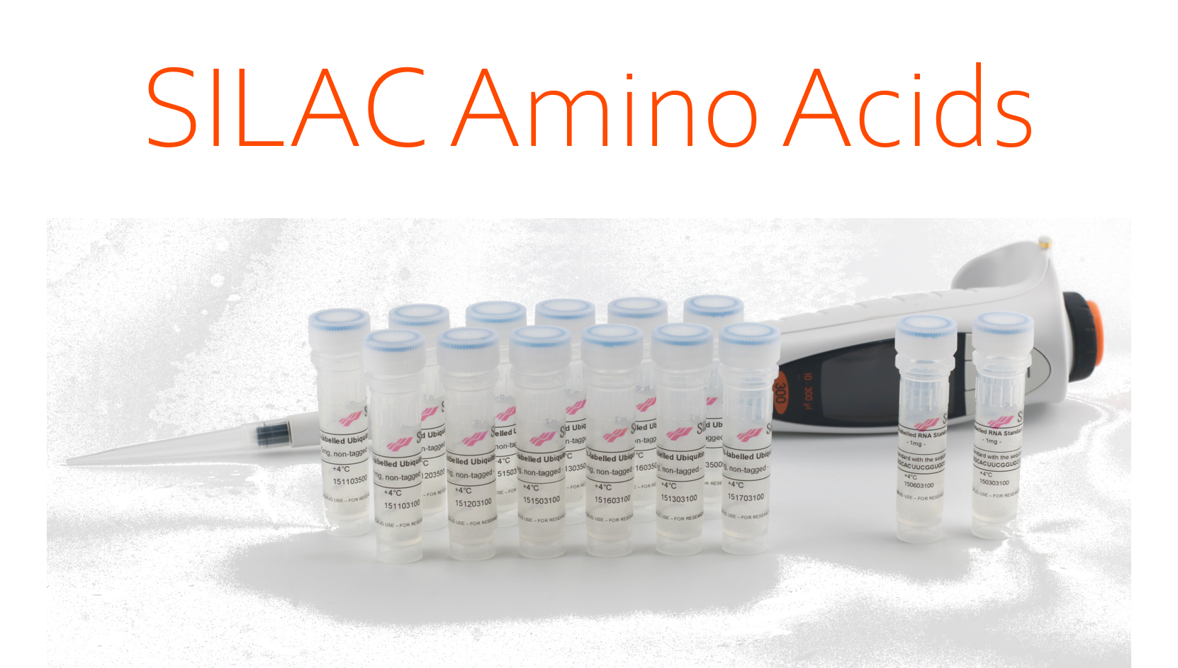
- SILAC DMEM, SILAC RPMI, SILAC dialyzed FBS and SILAC amino acids as single components.
The in-vitro SILAC workflow:
Figure 1 shows the workflow of the in-vitro SILAC procedure to quantitatively determine differences in the protein pattern of two cultures:
Step 1: Culture A (“light”) is supplemented with unlabelled amino acids, whereby culture B (“heavy”) is supplemented with labelled amino acids. As an example, in culture B, the 12C6-lysine is substituted by 13C6-lysine.
Step 2: Cells from both cultures are mixed in a 1:1 ratio. The proteins are isolated and digested with Lys-C, a protease which specifically cleaves at lysines.
Step 3: The proteolytic cleavage creates corresponding pairs of peptides stemming from culture A and B, differing by a molecular weight of 6 Da due to the molecular weight difference of the terminal 13C6-lysine. The ratio of the amount of “light” and “heavy” peptides is determined by mass spectrometry.
Silantes Components for in-vitro SILAC Experiments
Silantes offers all components that are necessary for a SILAC experiment. Each component is in a prepared sterile solution and ready for use. The components are available as individual products or in a kit.
Each kit consists of:
- 2 х 500 mL Silantes SILAC DMEM or RPMI media free of the amino acids lysine and arginine
- 2 х 50 mL Silantes dialyzed FBS
- Unlabelled L-lysine and L-arginine
- SILAC amino acids L-lysine and L-arginine
Please see the product numbers for all kits depending on the amino acid combinations in the table below.
High Quality of Silantes SILAC Components
The SILAC amino acids are available in all isotopic combinations. We guarantee an isotopic enrichment of > 98 atom % with a chemical purity of > 95 %. The isotopic purity is tested by mass spectrometry, whereas the chemical purity is tested by HPLC.
Figure 2 shows the growth kinetics of a model mammalian cell line (MDCK cells) using Silantes SILAC media and different labelling patterns of the SILAC L-lysines.
The experiment shows that the cells grow well on the Silantes SILAC components.
Figure 3 shows the incorporation of 13C6-lysine in an actin peptide (molecular mass = 586 Da) during the preparation of the "heavy" culture for a SILAC experiment.
A comparison of the 586 Da peak at t = 0 hours stemming from the unlabelled actin peptide and the 589 Da peak at t = 8 days stemming from the corresponding labelled actin peptide indicates that the cell culture is fully labeled after 8 days (4 passages).
That the nominal difference of the peaks is 3 Da (and not 6 Da) is due to the fact that the ratio m/z (x-coordinate) is 2 (instead of 1).
[1] Ong, S.E., Blagoev, B., Kratchmarova, I., Kristensen, D.B., Steen, H., Pandey A., and Mann, M. (2002). Stable isotope labeling by amino acids in cell culture, SILAC, as a simple and accurate approach to expression proteomics, Mol.Cell. Proteomics 1, 376–386.
|
|
|
|
|
|
|
|
|

